Figure 7.
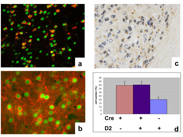
Cre expression in striatal neurons. (a) Retrograde labeling of striatonigral neurons within the striatum (red fluorescence). Cre was labeled by immunofluorescence (green fluorescence). (b) Double immunofluorescence against Cre (green fluorescence) and neuron-specific class III tubulin (TujI monoclonal antibody, red fluorescence). (c) Expression of Cre protein in neurons expressing D2R mRNA by in situ hybridization with a riboprobe specific for D2R mRNA (blue) followed by immunohistochemistry with Cre specific antibody (brown). (d) Quantification of Cre positive neurons expressing D2R mRNA. Neurons expressing either Cre protein or D2R mRNA were also counted. Reported is the % of neurons per each group. The data are expressed as mean ± SEM. a-b-c are coronal sections; (a,b 400×; (b) 1000×.

