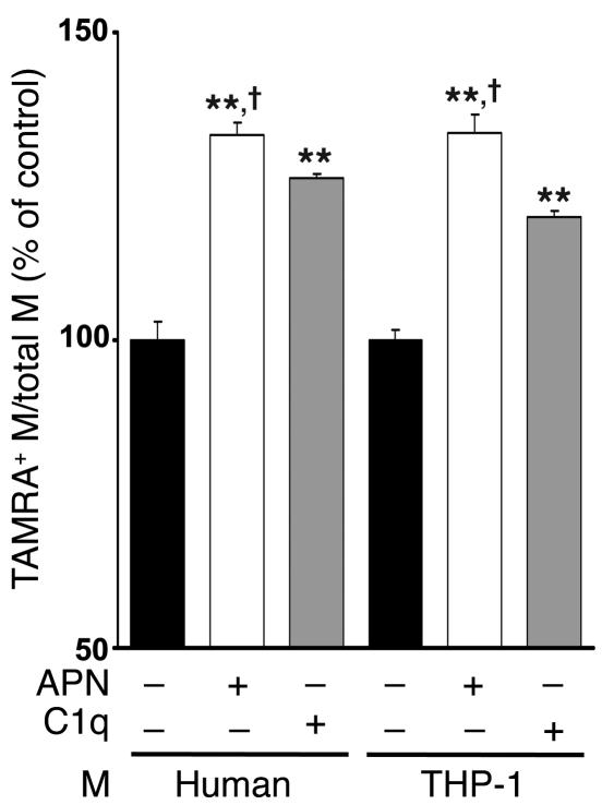
Figure 5. Adiponectin promotes the phagocytosis of apoptotic bodies by macrophages in vitro.
Apoptotic Jurkat T cells were preincubated for 1 hour with recombinant adiponectin from baculovirus-insect (APN) (50 μg/ml), human C1q (50 μg/ml), or vehicle. Macrophages were then incubated for 30 minutes with TAMRA, SE–labeled Jurkat cells that were either viable or apoptotic due to UVB exposure. Upon mixing apoptotic cells with macrophages, adiponectin and C1q were diluted to a final concentration of 10 μg/ml. Phagocytosis was assessed by flow cytometry. Macrophages were stained with FITC-conjugated anti-human macrophage antibody, and the percentage of phagocytic macrophages was calculated as TAMRA, SE–positive (+) macrophages/total macrophages × 100%. Phagocytic macrophages of control were 32.8% ± 1.0% (human) and 23.6% ± 0.4% (THP-1). **P < 0.01 versus vehicle; †P < 0.05 versus C1q (n = 6–7).

