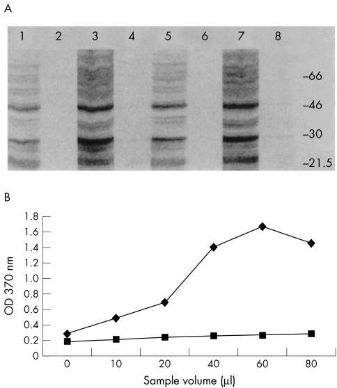
Figure 4.
(A) An immunostained western blot of the SDS extracted proteins from RPE and MCF7 cell matrices and 10-fold concentrated media fractions. Lane 1, RPE RAdTIMP-3 media; lane 2, RPE RAdlacZ media; lane 3, RPE RAdTIMP-3 matrix; lane 4, RPE RAdlacZ matrix; lane 5, MCF7 RAdTIMP-3 media; lane 6, MCF7 RAdlacZ media; lane 7, MCF7 RAdTIMP-3 matrix; lane 8, MCF7 RAdTIMP-3 media. (B) A line chart showing the results of ELISA detection of endogenous TIMP-3 in established RPE (upper line) and keratocyte (lower line) cell cultures. The control cells show relatively little endogenous TIMP-3 production compared with RPE cells.

