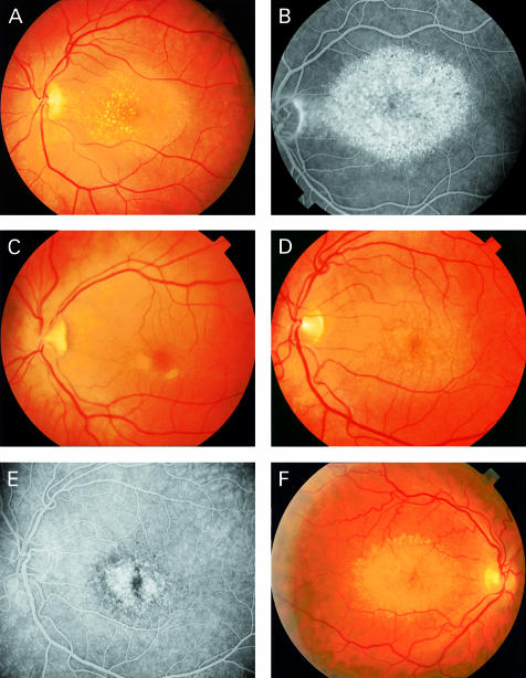
Figure 3.
Funduscopic pictures of CACD/dominant drusen patients. Family A: Patient III-10 (A) at age 50 demonstrates drusen with central localisation, the geographic area of atrophy of RPE and choriocapillaris is clearly visible on the fluorescein angiogram (B). Patient IV-1 (C) shows parafoveal lesions; fluorescein angiography (not shown) demonstrated loss of choriocapillaris and RPE typical for early CACD. Patient IV-8 (D) demonstrates a clinical picture at age 42 which resembles that of his mother (III-9) and aunt (III-10), the fluorescein angriogram shows central atrophy (E). Family B: in patient III-8 (F) the drusen are located mainly at the peripheral border of the associated CACD (age 51).

