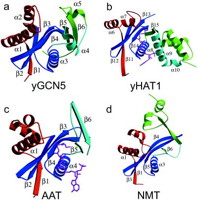
Figure 1.
Structure of yGCN5 and related enzymes. (a) Structure of the yGCN5 HAT domain. The four subdomains of the protein are color-coded; the structurally conserved subdomain that makes up the core (motifs A and D) is colored blue, motif B is colored aqua, and the N-terminal and C-terminal flanking regions are colored red and green, respectively. (b) Structure of residues 130–320 (C terminus) of HAT1 with the same orientation and color coding as in a. The AcCoA cofactor is shown in magenta. (c) Structure of SmAAT with the same orientation and color coding as in a. The CoA cofactor is shown in magenta. (d) Structure of residues 80–260 (N terminus) of NMT with the same orientation and color coding as in a.

