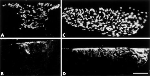
Figure 3.
Immunofluorescent localisation of tenascin (B, D) in sections of transgenic lenses from P21 mice (A, B) or rat lenses cultured for 4 days with TGFβ (C, D). Sections are counterstained with Hoechst dye (A, C). In all plaques, tenascin is predominantly expressed in cells that are closely associated with the lens capsule (B, D). As cells are displaced from the lens capsule, reactivity for tenascin is gradually lost (B, D). Scale bar: 80 μm.

