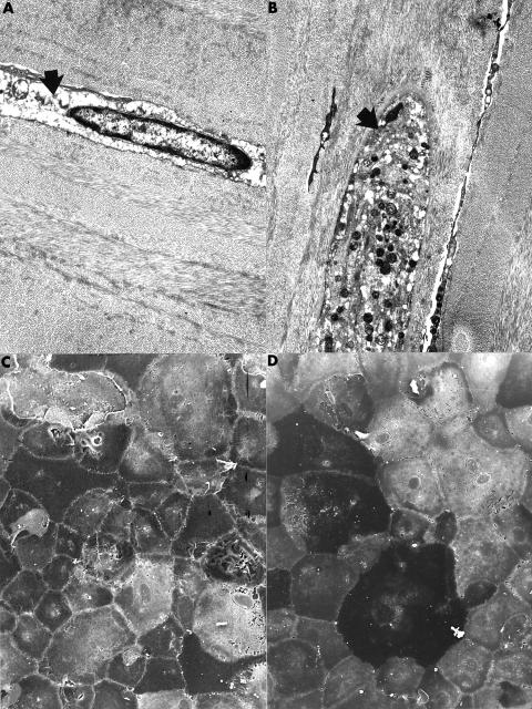
Figure 1.
Transmission electron micrographs of the corneal stroma showing representative abnormal keratocytes. One keratocyte has a markedly electron lucent cytoplasm (A from case 14) (arrow); in the other (B) the cytoplasm is distended and contains numerous electron dense bodies (arrow). (A) ×6500; (B) ×5100. Scanning electron micrograph of the corneal endothelium of case 14 (C) and case 15 (D) showing abnormal endothelial cells with a marked variation in shape and size covering a representative portion of Descemet's membrane. (C) ×400; (D) ×350.

