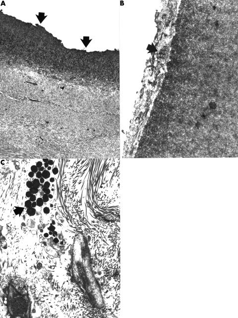
Figure 2.
Transmission electron micrographs of the posterior cornea of case 14 (A) and case 15 (B) showing abnormal Descemet's membranes. Part of the Descemet's membrane in case 14 had an uneven thickness and ragged posterior surface that lacked endothelial cells (A) (arrows). The posterior surface of regions of Descemet's membrane in case 15 was covered with a collagenous layer that contained prominent foci of broad banded collagen (arrow). (A) ×7000; (B) ×16 000. (C) Transmission electron micrograph of iris from case 14 showing a cluster of melanosomes within the iris stroma. In contrast with the normal iris these granules are not apparent within the cytoplasm of a melanocyte (arrow) (×5100).

