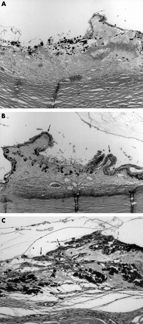
Figure 1.
(A) Low power photomicrograph of pars plicata showing severe, total destruction of ciliary processes with pigment clumping, loss of vessels, and little epithelial regeneration in case 5, 7 months after diode treatment. Haematoxylin and eosin. Original magnification ×40. (B) Low power photomicrograph of pars plicata showing damaged central zone with sparing of anterior and posterior processes (arrows) in case 5, 7 months after diode treatment. Haematoxylin and eosin. Original magnification ×40. (C) Medium power photomicrograph of haphazardly regenerated non-pigmented epithelium (arrows) over damaged pars plicata with underlying pigment clumping in case 3, 12 months after diode treatment. Haematoxylin and eosin. Original magnification ×100.

