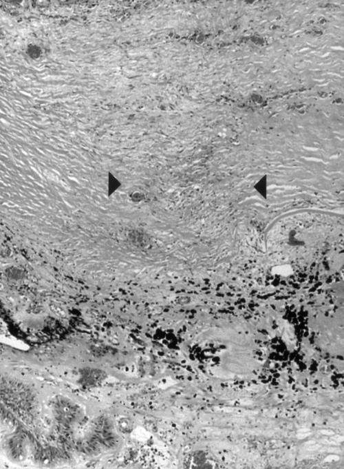
Figure 4.
Low power photomicrograph of scarred sclera (between arrowheads) with increased density of fibroblasts and new vessels overlying severely damaged pars plicata with total destruction of ciliary processes and pigment clumping in case 6, 4 months after diode treatment. Haematoxylin and eosin. Original magnification ×40.

