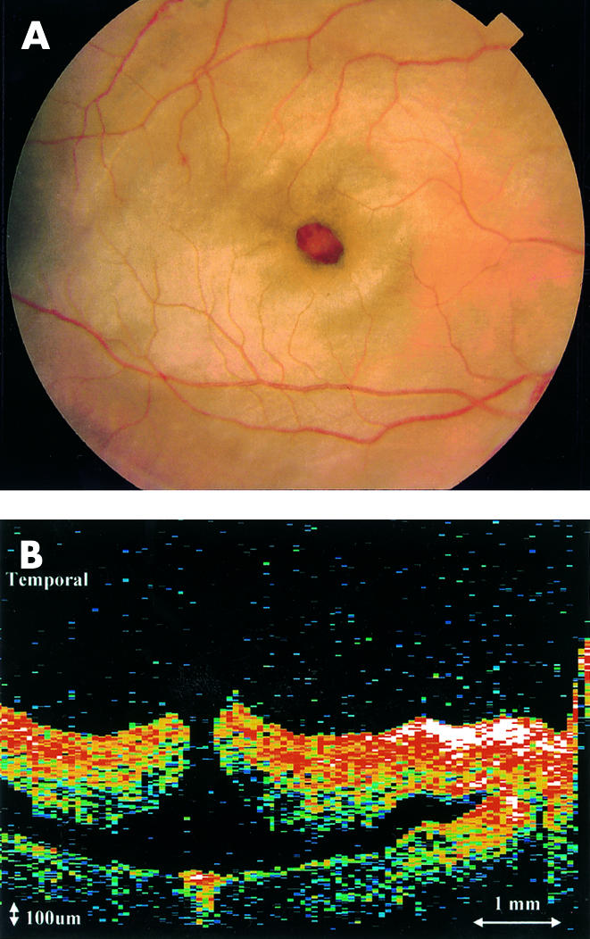
Figure 1.

(A) Right macula of 15 year old boy with extensive commotio retinae over posterior pole and an associated macular hole at 1 day after blunt injury. (B) Horizontal OCT scan through centre of macula confirms a full thickness macular hole and demonstrates extensive disruption of photoreceptor outer segment/retinal pigment epithelium layer. The optic disc is seen at the nasal edge of the scan.
