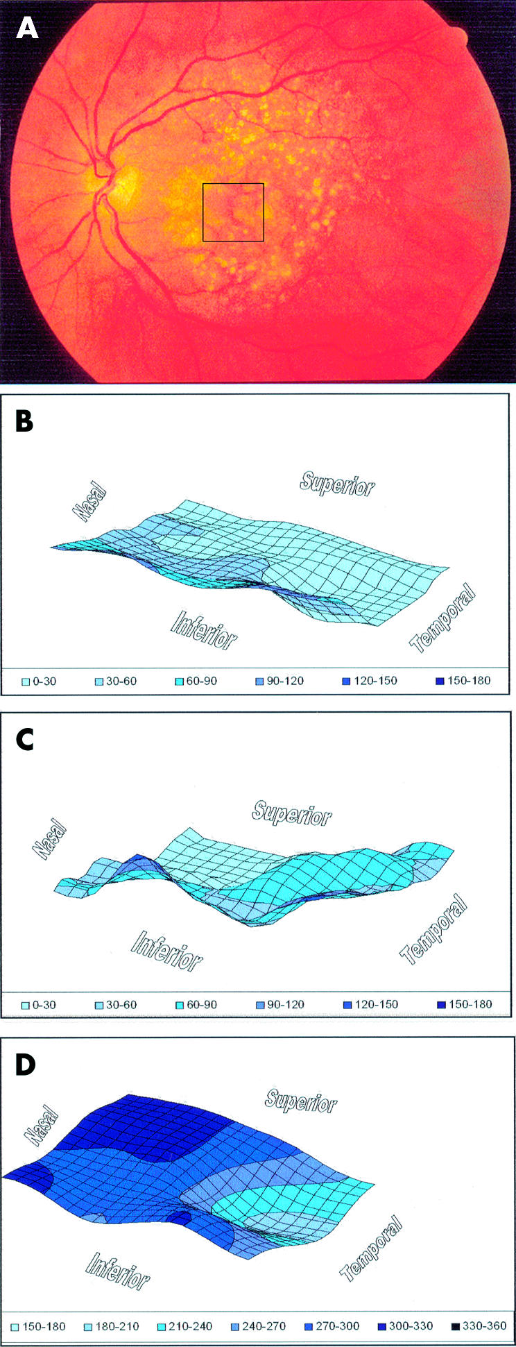
Figure 1.

Imaging was performed in the left eye of a patient with atrophic age related macular degeneration. (A) Optical section images were obtained from a scan through the retinal area indicated by the box overlaid on the fundus photograph. Optical section images were analysed to generate three dimensional topographic maps of the (B) vitreoretinal surface, (C) chorioretinal surface, and (D) retinal thickness. The relative surface height and retinal thickness are indicated in μm and pseudocolour coded for display. The vitreoretinal surface displayed a depression corresponding to the area of reduced thickness. The chorioretinal surface showed topographic variations.
