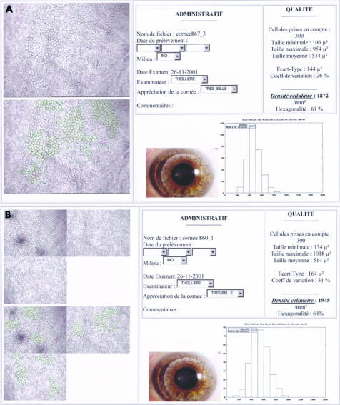
Figure 2.
Final result of analyses in mono-image (A) and tri-image (B) modes, sent by the cornea bank to the surgeon. This document, saved in HTML format, can be remotely transmitted. It contained all quantitative data (ECD, cell count) and morphometric data (area variation coefficient, min/max/mean/standard deviation of cell area, histogram of relative frequency distribution of cell areas, percentage of hexagonality). The one or three non-analysed and analysed video images of the endothelium are also printed, as is a non-specified image (in our case, an example of a graft). The technician also had a text box to note any comments.

