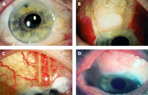
Figure 1.
(A) Pretreatment photograph of case 6 showing 360 degrees of conjunctival chemosis persisting 1038 days following filtration surgery. (B) Photograph of the same eye 1 day after injection of subconjunctival blood and placement of compression sutures. The IOP was 16 mm Hg. No blood has entered the filtration bleb or anterior chamber. The eye has been tilted inferiorly and the pupil is slightly more dilated, obscuring the peripheral iridotomy in this view. (C) Photograph of case 2 after blood injection. Conjunctival chemosis originated at the nasal aspect of the bleb, whereas the temporal aspect was flat. A single compression suture was therefore used. Note the conjunctival injection underlying the compression suture. The highest IOP for this eye was 20 mm Hg. (D) Final review photograph of case 5 showing linear conjunctival scarring and flattening of the conjunctiva after compression sutures and autologous blood. The compression sutures have been removed.

