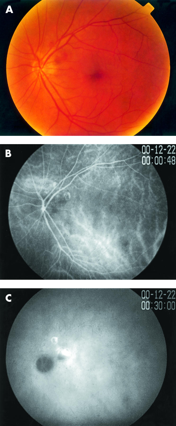
Figure 2.

Left eye. (A) Fundus photograph showing two haemorrhagic pigment epithelial detachments with underlying choroidal polyps in the peripapillary region. (B) ICGA (early phase, 0 minutes 48 seconds) showing early hyperfluorescent of the polypoidal lesions. (C) ICGA (late phase, 30 minutes 1 second) showing the classic ring-like silhouette staining of the polyps.
