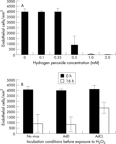
Figure 4.
Changes in corneal endothelial cell (EC) density following exposure to H2O2.. EC density was quantified by laser scanning confocal microscopy. Mean values (SD) from triplicate specimens in one representative experiment are shown. (A) Concentration dependent damage to EC following incubation of corneas in H2O2 at concentrations ranging from 0.1 mM to 2.0 mM for 16 hours. (B) Viability of EC was determined with scanning laser confocal microscopy in AdCL infected (2.5 × 10⁷ pfu), Ad0 (2.5 × 10⁷ pfu), and mock infected corneal specimens. After 16 hours of incubation in 0.5 mM H2O2, significantly more EC were viable in the AdCL transduced group.

