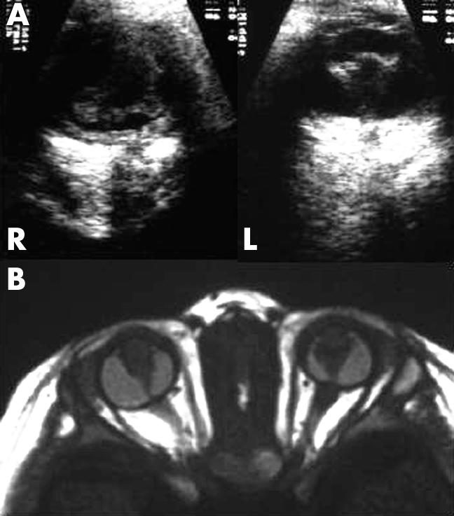
Figure 1.

Ocular findings in patient 2. (A) R = right eye; L = left eye. Ultrasonography suggests a funnel-shaped retrolental mass in each eye with shortened axial lengths. (B) T1 weighted magnetic resonance imaging clarifies microphthalmic eyes, retrolental masses, abnormal lenses, and enhancement in the anterior chambers due to elongated ciliary processes.
