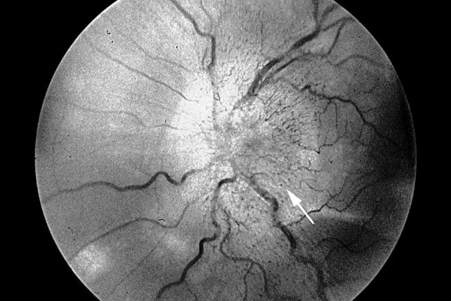
Figure 1.

Fundus photograph of the left optic disc shows diffuse swelling and increased surface vascularity. Additionally, a more focal elevation at the inferotemporal quadrant gives a nodule-like appearance to the disc swelling (arrow). The irregular white spot at the lower left corner of the figure is a photographic artefact.
