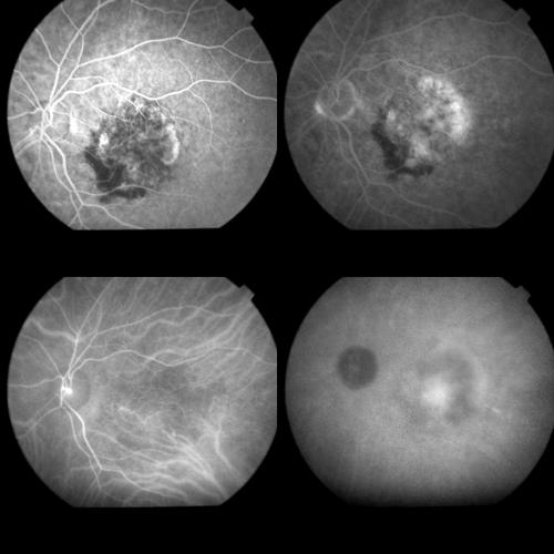
Figure 10.
Top left: FA image (59 seconds) of minimally classic AMD related CNV. Top right: Late FA phases (8 minutes) disclosing the extension of the occult and the classic components of the lesion. Bottom left: Early ICGA phase (1 minute) showing the filling of the CNV net. Bottom right: Late ICGA phases (42 minutes) revealing the staining of both the component of the CNV.

