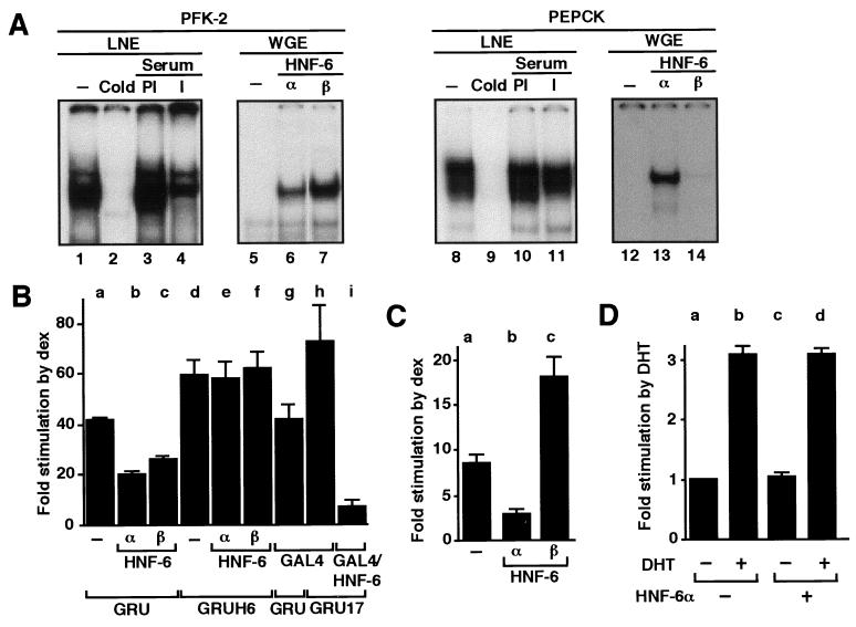
Figure 1.
Binding of HNF-6 to the GRU inhibits glucocorticoid stimulation of the pfk-2 and pepck genes. (A) Radiolabeled oligonucleotide probes containing the HNF-6-binding sites from the pfk-2 or pepck GRU were incubated with liver nuclear extracts (LNE) or with wheat germ extracts (WGE) programmed or not to synthesize HNF-6α or β. A 50-fold excess of cold probe, the serum of a rabbit immunized (I) against recombinant HNF-6, or preimmune serum (PI) was added as indicated. For lanes 1 and 8, no competitor or serum was added; for lanes 5 and 12, unprogrammed extracts were used. (B) FTO-2B cells were transiently cotransfected with expression vectors for the recombinant proteins indicated and with a luciferase reporter construct driven by the pfk-2 promoter linked to the intact pfk-2 GRU (GRU) or to the GRU in which the HNF-6-binding site has been disrupted (GRUH6) or replaced by a GAL4-binding site (GRU17). After transfection, the cells were treated for 24 h with dexamethasone and assayed for luciferase activity as described (10, 15). (C) H4IIE cells were treated as in B, except that the reporter construct was driven by the pepck promoter and the cells were exposed for 16 h to dexamethasone. (D) FTO-2B cells were transiently transfected with a luciferase reporter construct driven by the pfk-2 promoter linked to the intact pfk-2 GRU and with expression vectors for the androgen receptor and for HNF-6α as indicated. After transfection, the cells were treated for 24 h with dihydrotestosterone (DHT) as indicated. Values are means ± SEM from three to five experiments.

