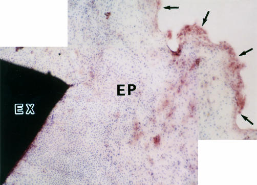
Figure 2.
Immunocytochemical study of ICAM-1 expression on HCE cells. HCE cells of epithelial outgrowth from the limbal explant after 7 days of incubation were stained with anti-ICAM-1 mAb by the ABC method, visualised with AEC, and counterstained with haematoxylin. Positive staining for ICAM-1 (brown stain) was found predominantly on HCE cells in the leading zone. Original magnification, × 34. Limbal explant (EX). Epithelial outgrowth (EP). Leading edge of epithelial outgrowth (arrows).

