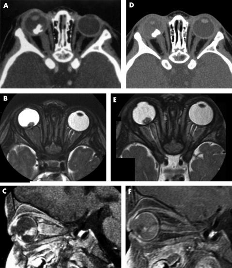
Figure 1.
(A–C) Left unilateral retinoblastoma with orbital optic nerve involvement at diagnosis. Axial post-contrast CT scan (A), axial T2 weighted (B), and enhanced fat saturated T1 weighted sagittal (C) MR images. Intraocular calcified tumour with low signal intensity on the T2 weighted sequence and high signal intensity after gadolinium injection on the T1 weighted sequence. The optic nerve is enlarged and shows the same signal abnormalities as the primitive intraocular tumour. (D–F) Preoperative imaging after two courses of etoposide and carboplatin. Axial post-contrast CT scan (D), axial T2 weighted (E), and enhanced fat saturated T1 weighted sagittal (F) MR images. The intraocular tumour volume remains unchanged but post-contrast MR (F) shows a decreasing enhancement. Optic nerve involvement has significantly decreased, with only slight residual enlargement and enhancement.

