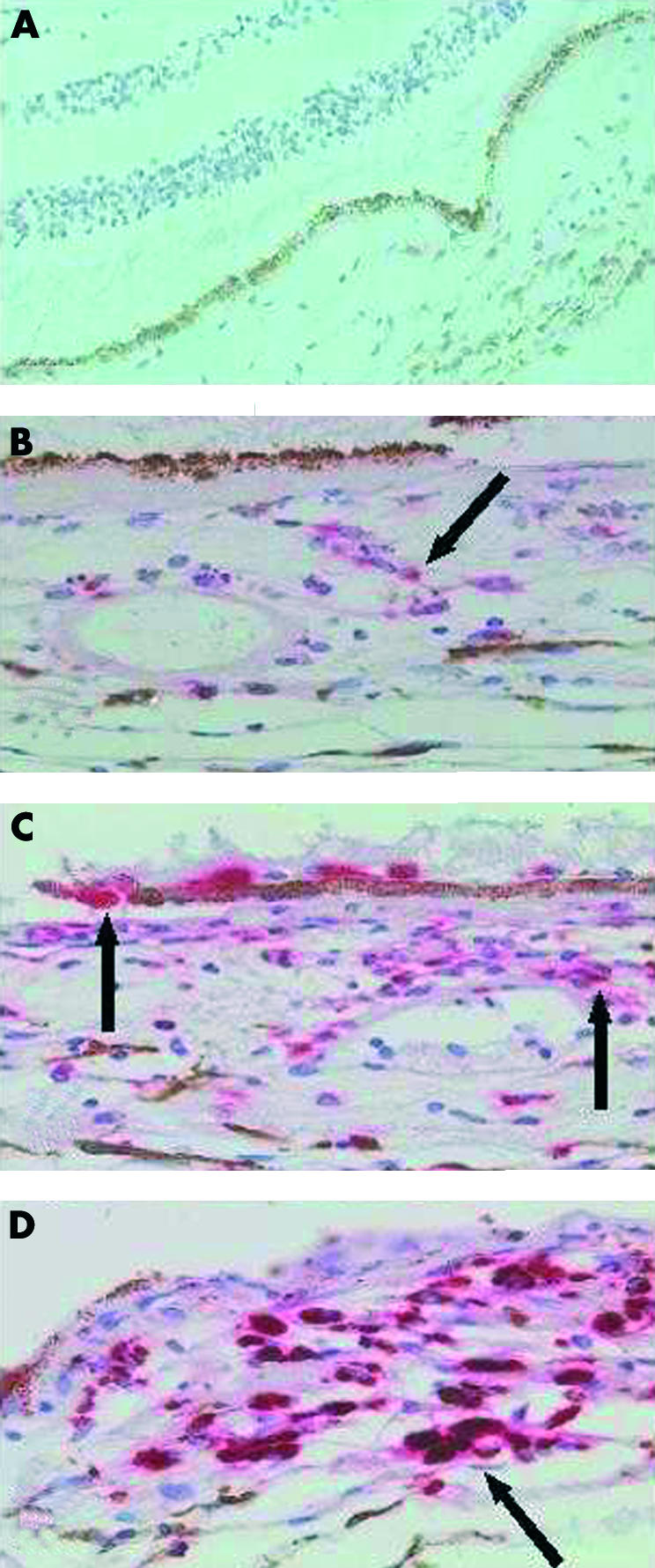
Figure 3.

Pattern of CD68 staining in normal and laser treated retinas. Haematoxylin and eosin and anti-CD68 antibody. (A) Normal retina with no CD68 staining in the choroid, 50× magnification. (B) CD68 staining in the choroid 1 day post-laser, 100× magnification. (C) CD68 staining 2 days post-laser. Note CD68 positive cells associated with RPE and perivascular cuff, 100× magnification. (D) CD68 staining 6 days post-laser showing more intense staining compared to days 1 and 2, 100× magnification.
