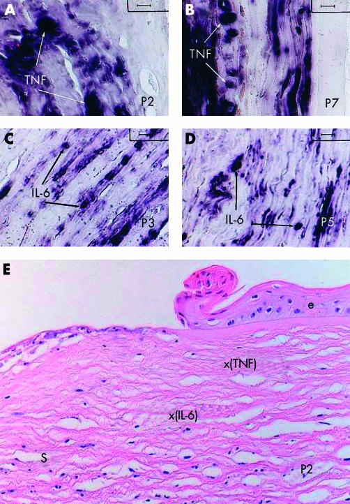
Figure 1.
Detection of TNF-α (A, B, and E) and IL-6 (C, D, and E) mRNA by non-radioactive in situ hybridisation in corneal sections from patients with corneal ulcerations and/or perforations associated with rheumatoid arthritis. (A) Representative TNF-α mRNA hybridisation pattern of the corneal keratocytes in a paraffin section from patient 2 (hybridisation score: +). (B) Representative TNF-α mRNA hybridisation pattern of the corneal keratocytes in a paraffin section from patient 7 (hybridisation score: ++). (C) Representative IL-6 mRNA hybridisation pattern of the corneal keratocytes in a paraffin section from patient 3 (hybridisation score: ++). (D) Representative IL-6 mRNA hybridisation pattern of the corneal keratocytes in a paraffin section from patient 5 (hybridisation score: +). (E) Representative corneal section from patient 2, stained with haematoxylin and eosin showing the relative position of the stroma cells yielding positive mRNA hybridisations either for TNF-α (x(TNF)), or for IL-6 (x(IL-6)). A positive reaction is indicated by a dark blue-violet colour as a result of the corresponding reaction using the digoxigenin detection kit from Boehringer-Mannheim. TNF = TNF-α, e = corneal epithelium, s = corneal stroma (original magnification (A–D) ×1000, (E) ×200).

