Figure 1.
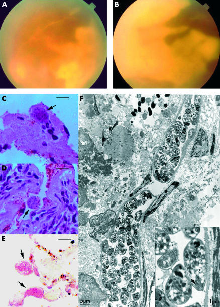
(A) Fundus appearance of the left eye. (B) Fundus appearance of the right eye. (C) Light micrograph shows necrotic retina with occasional inflammatory cells and Toxoplasma cyst (arrow). (D) Light micrograph shows the necrotic retinal pigment epithelium with Toxoplasma cysts (arrow) and the choroid with a dense infiltrate of lymphocyte and plasma cells (haematoxylin and eosin, bar = 10 μm). (E) Immunohistochemistry with alkaline phosphatase. Two Toxoplasma cysts (arrows) positively dyed pink (bar = 10 μm). (F) Electron micrograph of a paraffin embedded tissue post processed for electron microscopy of the chorioretinal biopsy shows two Toxoplasma cysts with micronemes (inset, arrow) and white spherules with hazy borders amylopectin granules (inset, asterisk). The inset shows higher magnification of a bradizoite, showing typical apical structures. Magnification is represented in standard bars.

