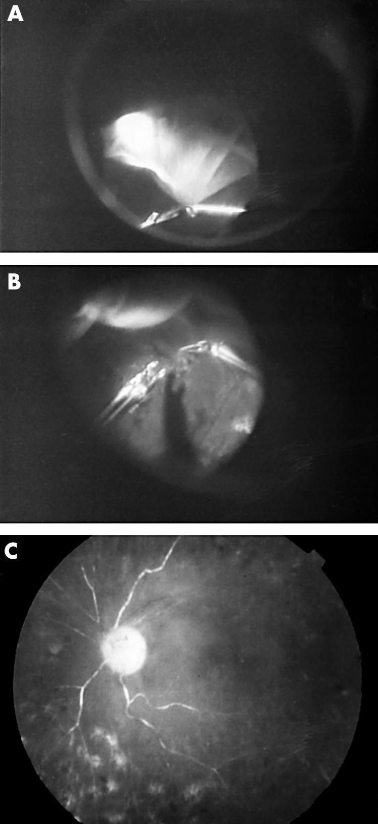
Figure 2.

Fundus photographs and intraoperative findings of the left eye of patient 3. The preoperative fundus was not visible because of dense vitreous haemorrhage. After removing the vitreous haemorrhage, tractional retinal detachment involving the macula was seen. (A) After relieving the tight adhesions at the periphery, a bullous retinal detachment can be seen. Then, PFCL was injected to stabilise the detached retina. (B) Thereafter, continued peeling and shaving the peripheral vitreous was performed. (C) Postoperative fundus. Retina is reattached; however, the visual acuity remained at light perception because a retinal artery occlusion occurred 2 months postoperatively.
