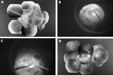
Figure 3.
Fundus photographs and intraoperative findings of the left eye of patient 15. (A) Preoperative fundus. Combined traction/rhegmatogenous retinal detachment can be seen. (B) Intraoperative findings. PFCL was injected into the centre of the funnel to flatten the detached retina. (C) The fibrovascular membranes on the stabilised retina were dissected under good visibility. (D) Postoperative fundus. Retina is reattached with a visual acuity of finger counting.

