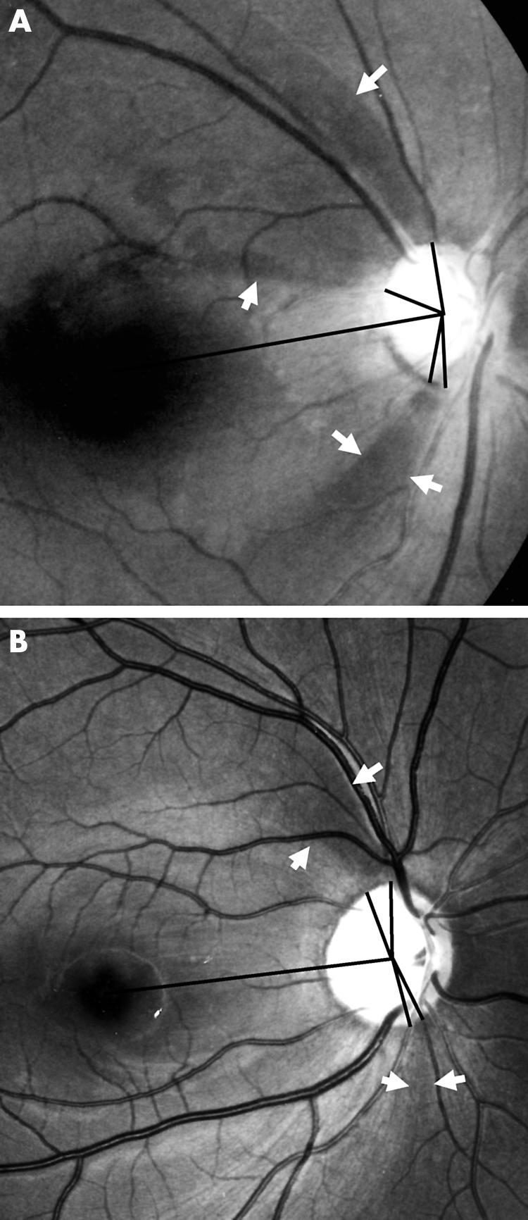
Figure 2.

Typical retinal nerve fibre layer (RNFL) photographs of normal tension glaucoma (NTG) (A) and primary open angle glaucoma (POAG) (B). (A) Localised NFL defects of NTG located in both superior and inferior retina are wider and closer to the fovea than those of POAG (B). (B) Localised NFL defects of POAG are far from the fovea and have very narrow widths.
