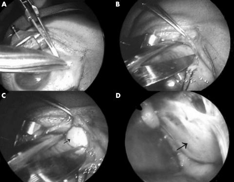
Figure 2.
(A) The first incision was made through the caruncle using Westcott scissors. (B) Gentle dissection with the tips of the scissors passed along the plane between Horner’s muscle and the medial orbital septum. (C) Retraction of the orbital contents with a malleable ribbon retractor resulted in exposure of the medial orbit and some mucoid material encountered (arrow). (D) The eggshell-like, rounded mucocele was shown (arrow).

