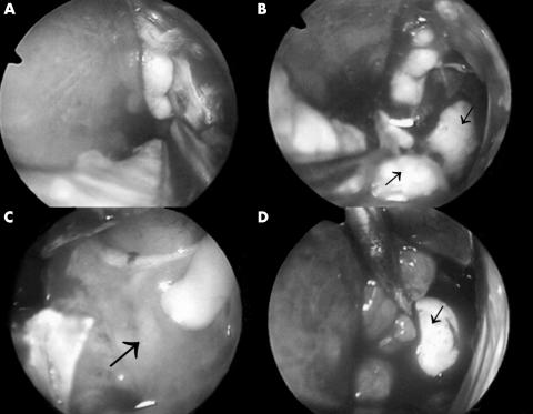
Figure 3.
(A) The outer wall of the mucocele was removed by forceps. (B) Yellowish mucoid material (arrows) was found after removal of outer shell of the mucocele. (C) The inner wall of the mucocele was shown (arrow). (D) A rubber catheter with a balloon (arrow) was inflated after excision of the mucocele.

