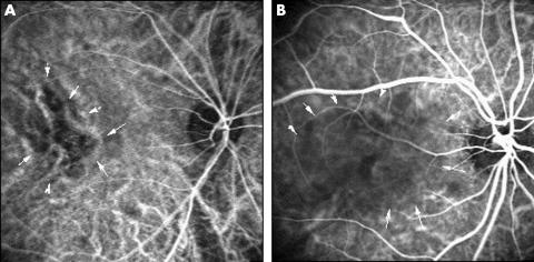
Figure 1.
Typical cases of delayed arterial filling using ICG angiography. (A) Right eye of a 52 year old man with CSC. ICG angiography (angle, 30°) 24 seconds after dye injection shows delayed arterial filling in the foveal region (arrows). (B) Right eye of a 41 year old man. Delayed arterial filling is observed 33 seconds after dye injection (arrows) (angle, 30°).

