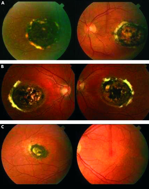
Figure 2.
The ocular phenotype. Colour fundus photographs: (A) individual II:1 a 67 year old female) showing centred on both foveae a well demarcated subfoveal area of chorioretinal atrophy with pigment hypertrophy and fibrosis at the edge and (B) her son (III:3, age 41 years), similar appearances both eyes; (C) her daughter (III:1, age 42 years) showing right eye, similar appearances; left eye, drusen-like deposits and retinal epithelial atrophy centred on the macula.

