
Figure 5.
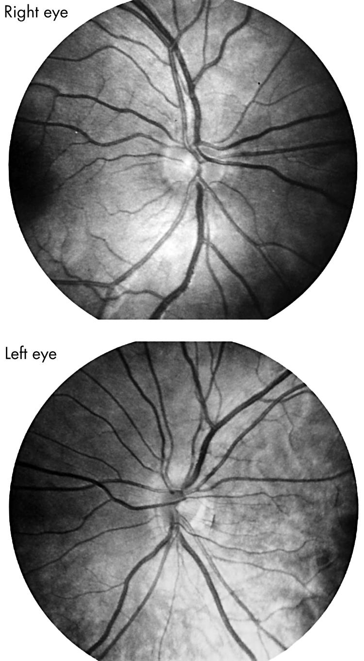
Fundus photographs of an 8 year old boy with unilateral high myopia due to segmental optic nerve hypoplasia left eye. Note that the inferotemporal portion of the left optic disc is truncated and separated from the adjacent pigment epithelium by a crescent of peripapillary tissue.