Figure 2.
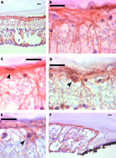
Nestin positive cells with an elongated morphology in the adult human retina. Composite of nestin immunocytochemistry of adult human retina to the show morphology, extent and distribution of elongated nestin positive cells with fibrous processes in the adult human retina. All scale bars represent 20 μm. (A) The peripheral retina contains large numbers of nestin positive cells, with elongated fibrous processes. Some are radially and others superficially arranged. This section of retina contains an area of cystic retinal change. (B) A high power view of the retinal surface in the peripheral retina. Radial and superficial fibres interlock and are continuous. (C) Some nestin positive cells form the innermost layer of the retina (arrowed). These cells give rise to processes running along the retinal surface. (D) In the anterior retina, some cells are found with speckled cytoplasmic staining (arrowed), in close association with nestin positive fibrous processes. (E) In the peripheral retina some speckled cells (arrowed) are seen to give rise to fibrous processes. (F) Nestin positive cells extend right up to the ora serrata. The cells with fibrous processes extend right up to the ora serrata.

