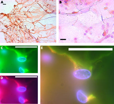
Figure 3.
Nestin positive cells in epiretinal membranes removed surgically, from the posterior retina. Composite of nestin positive cells in epiretinal membranes removed surgically. All scale bars represent 20 μm. (A) A clump of nestin positive cells, with elongated fibrous processes in an epiretinal membrane removed surgically. All nestin positive cells identified in epiretinal membranes (ERM) had elongated and fibrous processes. (B) A high power view of nestin positive cells in an ERM. Not all cells are nestin positive. Immunofluorescence demonstrates that GFAP (C, green) and nestin (D, red) co-localises in many cells in epiretinal membranes (E).

