
Figure 2.
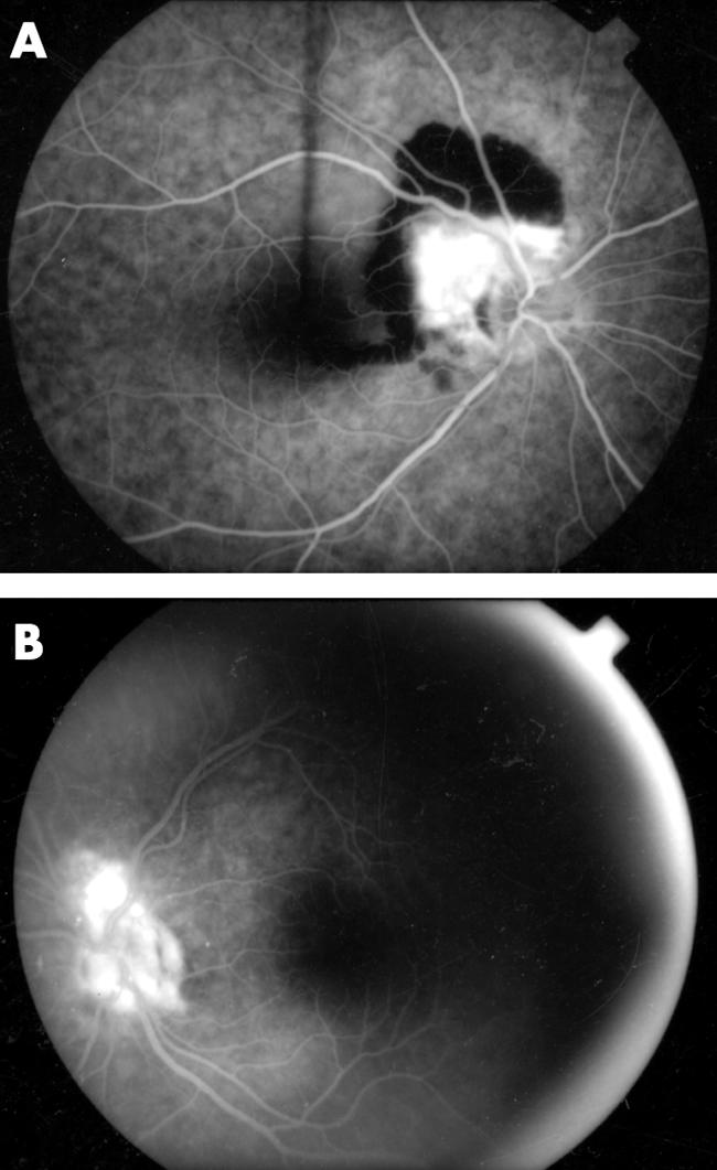
(A) Angiography of the right eye reveals a wedge shaped peripapillary CNV membrane. (B) A smaller CNV membrane is present angiographically superior to the left optic disc (late phase).
Official websites use .gov
A
.gov website belongs to an official
government organization in the United States.
Secure .gov websites use HTTPS
A lock (
) or https:// means you've safely
connected to the .gov website. Share sensitive
information only on official, secure websites.
(A) Angiography of the right eye reveals a wedge shaped peripapillary CNV membrane. (B) A smaller CNV membrane is present angiographically superior to the left optic disc (late phase).