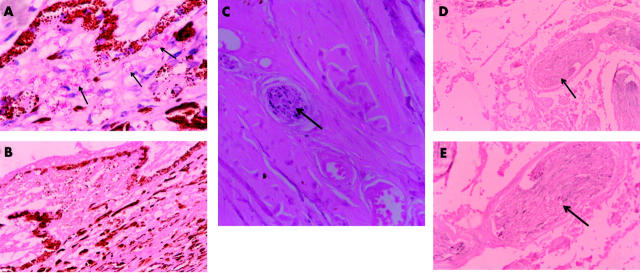
Figure 2.
Leprosy eyes, histopathology. (A) Photomicrograph showing ciliary process of the lepromatous eye infiltrated by macrophages containing Mycobacterium leprae (acid fast stain×400). (B) Photomicrograph of ciliary process showing complete absence of staining of ciliary nerve endings (immunohistochemical stain PGP 9.5×400). (C) Photomicrograph of sclera showing poor and patchy staining of a posterior ciliary nerve within the sclera (immunohistochemical stain PGP 9.5×400). (D) Photomicrograph of posterior ciliary nerve situated beside the optic nerve at its site of entry into the eyeball, showing complete absence of axonal staining (immunohistochemical stain PGP 9.5×400). (E) Magnified photomicrograph of posterior ciliary nerve situated beside the optic nerve at its site of entry into the eyeball, showing complete absence of axonal staining (immunohistochemical stain PGP 9.5×400).

