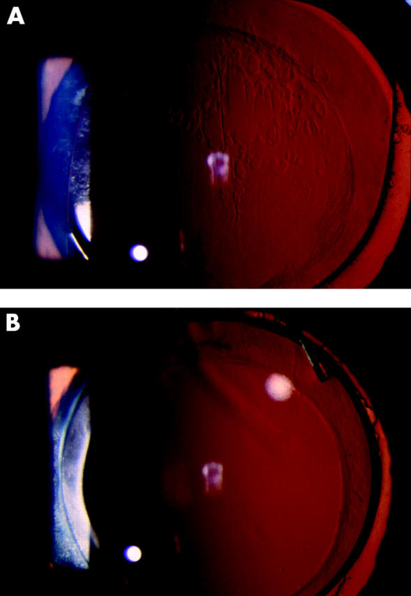
Figure 5.

Retroillumination photographs showing the bilateral eyes of a representative patient at 24 months after surgery. In an eye with a hydrogel IOL (A), fibrosis of the anterior capsule along the capsulorrhexis margin is marked. A flat proliferation of lens fibre cells over the posterior capsule is noted. However, swelling of these cells is slight. In the opposite eye with an acrylic IOL (B), fibrosis of the anterior capsule is slight and the posterior capsule is completely clear.
