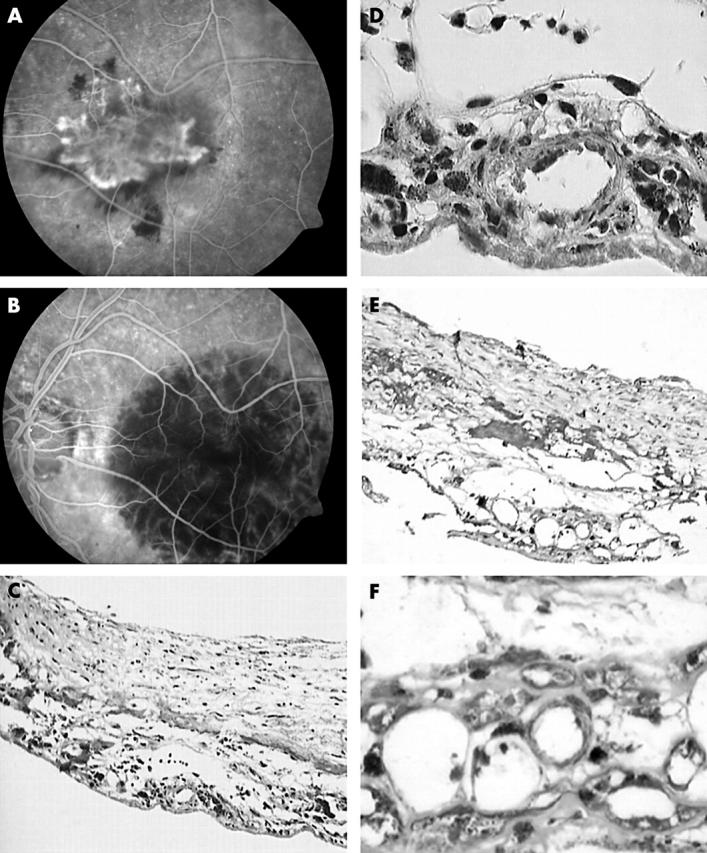
Figure 2.

(A) Early phase fluorescein angiography reveals subfoveal classic CNV. (B) Three days after PDT, early phase fluorescein angiography shows non-perfusion of the CNV and the treated area at the macula. (C) Overview of subretinally located fibrovascular tissue. Only a few vascular elements are retrieved even though the specimen is fairly rich in cells (Masson trichrome; original magnification ×20). (D) Detail of a capillary shows swollen endothelial nuclei and no occlusion of the lumen (Masson trichrome; original magnification ×100). (E) and (F) Fibrin deposition is found in the stroma of the CNV but not within the vessels. The vascular endothelium is severely damaged and a marginal polymorph nuclear cell can be seen. Some luminal debris is found but an occlusion is not seen ((E) PTAH; original magnification ×40 and (F) detail, PTAH; original magnification ×100).
