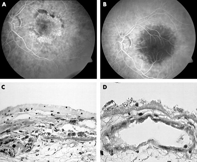
Figure 3.
(A) Fluorescein angiography shows well defined early bright hyperfluorescence consistent with a subfoveal classic CNV. (B) Three days after PDT, early phase of the fluorescein angiography reveals hypofluorescence at the macula. (C) Detail showing subretinal and intra-Bruch’s fibrovascular tissue separated by retinal pigment epithelium and diffuse drusen. A rare vascular element is suspected. Endothelial lining appears to be disintegrated (Masson trichrome; original magnification ×40). (D) Detail of a vein lined by abnormal endothelium. A thrombus formation is not seen (Masson trichrome; original magnification ×100).

