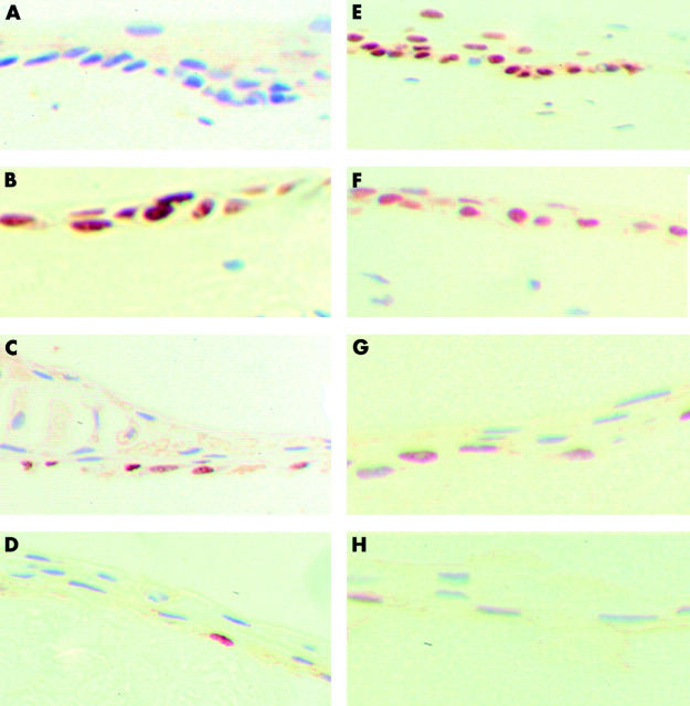
Figure 2.
Staining pattern of the explant with Ki67 and p63 on day 0, weeks 1, 2, and 3 (paraffin sections with diaminobenzidine tetrahydrochloride as chromogenic substrate; original magnification ×400). (A) The explant is negative for Ki67 on day 0. (B) At week 1 a significant increase in the expression of Ki67 in the basal and suprabasal epithelial cells is seen. (C) At week 2 only a few of the cells remain positive for Ki67. (D) By week 3 only an occasional cell in the basal layer is positive for Ki67. (E) On day 0 most of the basal cells are strongly positive for p63. (F) At week 1 most of the epithelial cells are positive for p63. (G) At week 2 a few of the basal cells remain positive for p63. (H) By week 3 only an occasional basal cell is positive for p63.

