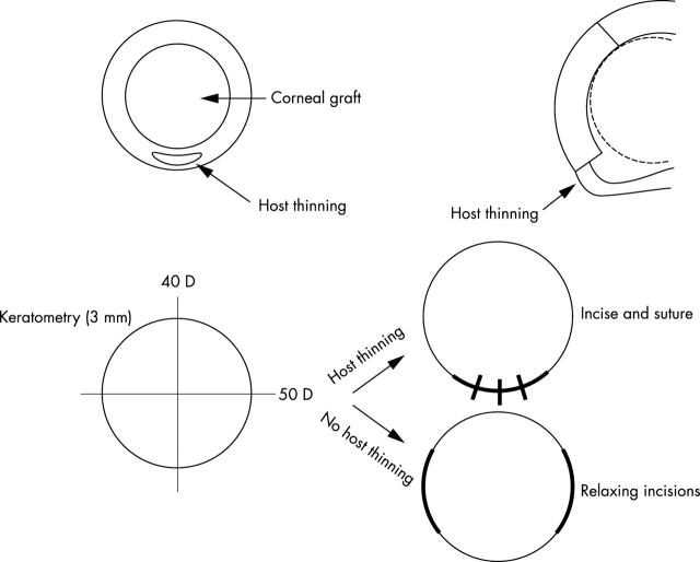
Figure 6.
Schematic diagram illustrating: (Top) progression of keratoconus in the inferior host cornea with inferior host thinning resulting in weakening of the inferior graft-host junction. (Bottom) The technique of compressive resuturing over the graft-host junction with host thinning and resultant flat meridian as opposed to relaxing incisions over the graft-host junction with the steep meridian.

