
Figure 1.
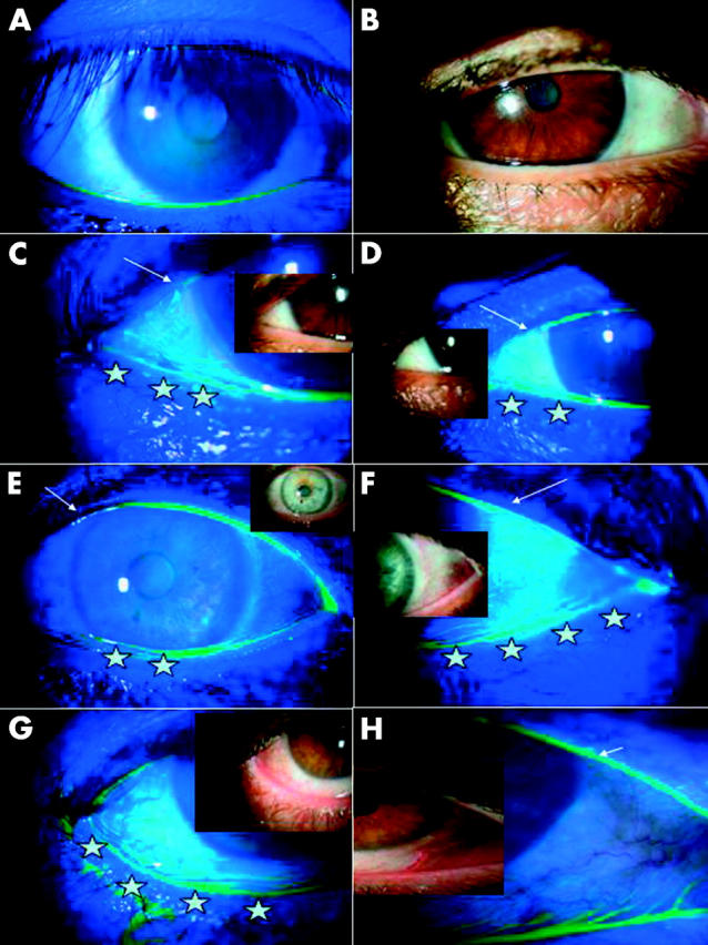
Fluorescein staining showing obliteration of tear meniscus of lower and upper lids by CCh. (A) In this representative ATD patient without CCh, there is continuous low tear meniscus of the lower and upper lids. (B to D) In this representative ATD with severe CCh, conjunctival folds were noted in the temporal bulbar conjunctiva (B). However, even if the meniscus was low, obliteration of the tear meniscus by temporal CCh becomes apparent when fluorescein is used, and is noted in both the lower and upper lids (C) and by nasal CCh (D) (stars and arrow respectively). (E and F) In this representative patient with severe CCh without ATD, obliteration of the tear meniscus of the lower lid (stars) and of the upper lid (arrows) by conjunctival folds is noted in both temporal and nasal bulbar conjunctiva, respectively. (G and H) Another representative patient with more severe CCh without ATD, obliteration of the tear meniscus of the lower lid (stars) and of the upper lid (arrows) by multiple conjunctival folds is noted in both temporal and nasal bulbar conjunctiva, respectively. Insets show the corresponding control without fluorescein.