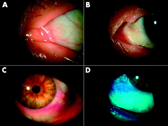
Figure 3.
Swollen puncta and rose bengal staining. (A) Normal appearance of lower and upper puncta in an ATD patient. (B) Swollen puncta with elongation and subcutaneous oedema of lower and upper puncta in a CCh patient. (C and D) Under normal light and a green filter, respectively, rose bengal staining is detected at non-exposure zone (inferior limbal region), pingueculae (exposure zone), and the mucosal surface of the inferior lid margin (due to CCh in the region) in a CCh patient without ATD.

