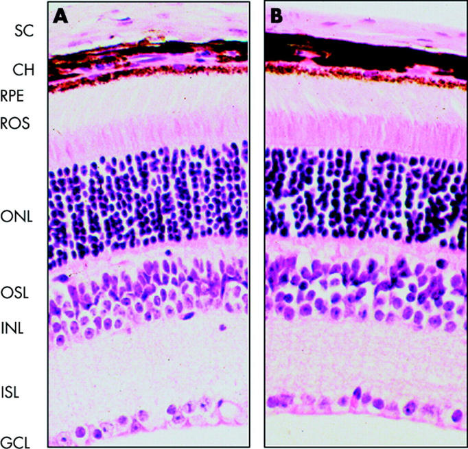
Figure 6.

Histochemical staining of T27aT15 mouse retina. Transverse paraffin sections (3 μm) from 3 month old wild type (A) and T27aT15 (B) mice were stained with haematoxilin and eosin, and visualised (magnification, ×250) as described under materials and methods. SC, sclera; CH, choroids; RPE, retinal pigment epithelium; ROS, photoreceptor segments of rods and cones; ONL, nuclei of rods and cones; OSL, outer synaptic layer; INL, neuron nuclear layer; ISL, inner synaptic layer; GCL, ganglion cell layer.
