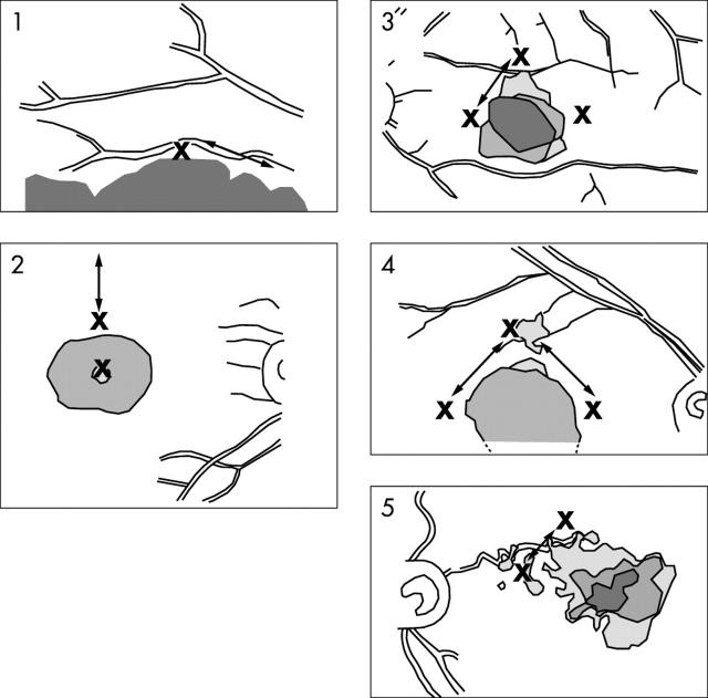
Figure 1.
Scotoma location in the five patients who reported the need to perform fixation related eye movements. The Xs indicate the position of the PRL previously identified during the reading task and the arrows the direction of the fixation related eye movements observed. For patients 1 and 2 the fixation related eye movements were observed to occur from the PRL to an area not included in the PRL identified during reading. For patients 3, 4, and 5 these movements were performed between two PRL areas. Illustrations represent the fundus as seen through the SLO. As a result, the image is vertically inverted relative to the projection of the scotoma in visual space.

