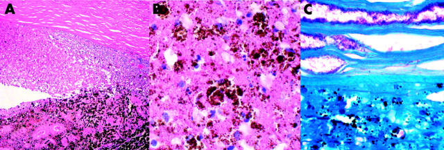
Figure 2.
(A) Haematoxylin and eosin stain reveals necrosis of iris and inflammatory exudates in the anterior chamber. (B) The anterior chamber exudate displays several melanophages and necrotic cells (haematoxylin and eosin). (C) Ziehl-Nielsen’s stain shows a myriad of acid fast bacilli in the deep corneal stroma and in the anterior chamber exudates.

