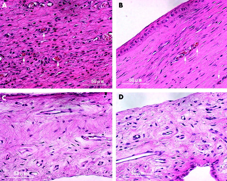
Figure 6.

Light micrograph of a cornea (top) and an iris (bottom) stained with haematoxylin eosin at day 0 (left) and day 3 (right). (A) Corneal neovascularisation is filled with thrombus (arrow) with no severe inflammatory changes. (B) Small vessels are packed with aggregated erythrocytes and platelets (arrow). No inflammatory changes are detected and there is a small cavity of neovascularisation. Iris vessels are patent and no morphologic changes are seen at day 0 (C) or day 3 (D) (haematoxylin eosin staining, original magnification ×400).
