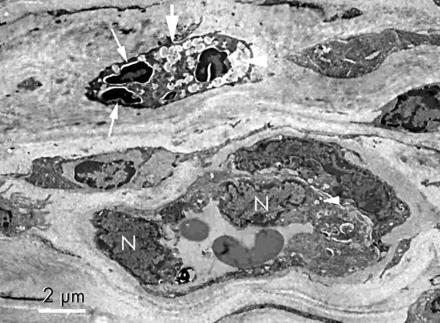
Figure 7.
Transmission electron micrograph of cornea on day 3 after photodynamic therapy. Cell remnants and some amorphous materials (large arrow) are packed in the vascular lumen (large arrow head). Nuclear alterations of endothelial cells (small arrow) are seen. Vacuolisation of the endothelial cell cytoplasm (small arrow head) and swelling of endothelial nuclei (N) are also observed (uranyl acetate and lead citrate ×2000).

