
Figure 4.
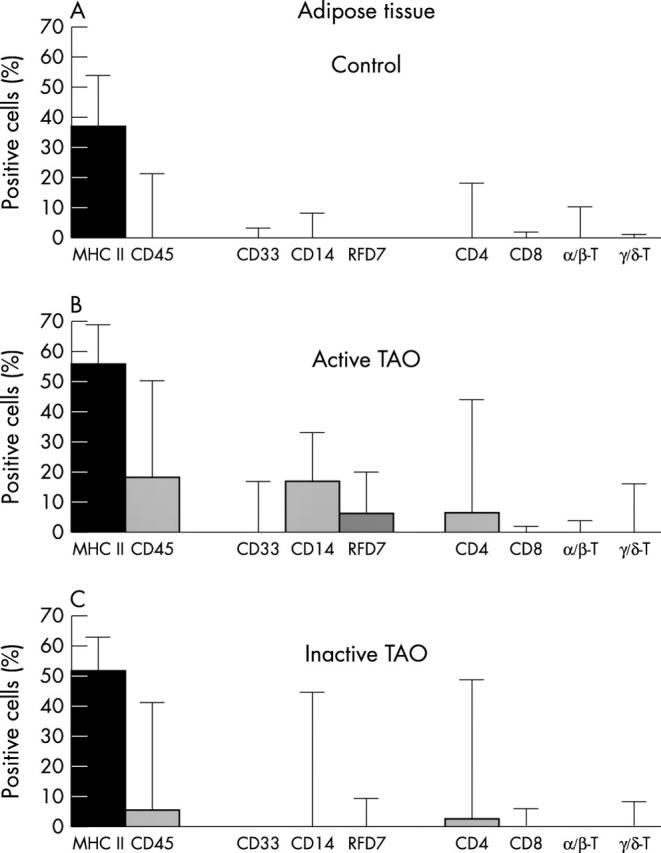
Cells in periorbital adipose tissue of (A) controls, (B) patients with A-TAO, and (C) patients with I-TAO. For markers and statistics notes, see legend to figure 3 . Control adipose tissue revealed lower leukocyte numbers than control fibrovascular tissue. This is in line with a much less pronounced infiltration of adipose tissue by disease associated myeloid and lymphoid cell types. Nevertheless, TAO associated alterations were qualitatively similar to those observed in the fibrovascular septae, with the important exception of freshly invading CD33+ blood monocytes. Also note that, in stark contrast to fibrovascular tissue, decreased disease activity was accompanied by a concomitant decrease in CD4+ cells.